Orthopedic Soft Tissue Research Program
We are a team of clinicians, scientists, and engineers who carry out research on the health and function of joints and surrounding tissues. Our research relates to the mechanisms of joint injuries commonly seen in active individuals, including injury to articular cartilage, tendon, ligament, meniscus, and intervertebral disc. We are developing targeted approaches that promote more robust and consistent tissue repair to restore joint mobility and prevent degeneration.
Research Team and Advisory Committee
Our program is comprised of a multidisciplinary team of investigators with expertise in cell biology, molecular biology, developmental biology, biomechanics and biomaterials, chemistry, and orthopedic surgery. A particular strength is the close collaboration between clinicians, bioengineers, and basic scientists, which facilitates clinically relevant research that can be translated to clinical applications.
Advisory Committee Members
The Advisory Committee of our program consists of senior scientists and clinician-scientists from HSS who have a history of working within our program. They leverage their experience and perspective in the field of orthopedics to strengthen the program by advising on overall grant writing strategies, by helping with career planning for early- to mid-level faculty, and by assisting with strategic planning for the program.
Madhu M. Bhargava, PhD
Mary Goldring, PhD
Jo A. Hannafin, MD, PhD
Peter Torzilli, PhD
Russell F. Warren, MD
Research Areas


Select Publications
- Hwang SM, Feigenson M, Begun DL, Shull LC, Culley KL, Otero M, Goldring MB, Ta LE, Kakar S, Bradley EW, Westendorf JJ. Phlpp inhibitors block pain and cartilage degradation associated with osteoarthritis. J Orthop Res. 2017 Oct 25. doi: 10.1002/jor.23781. [Epub ahead of print] PubMed PMID: 29068480.
- Holyoak DT, Otero M, Armar NS, Ziemian SN, Otto A, Cullinane D, Wright TM, Goldring SR, Goldring MB, van der Meulen MCH. Collagen XI mutation lowers susceptibility to load-induced cartilage damage in mice. J Orthop Res. 2017 Sep 12. doi: 10.1002/jor.23731. [Epub ahead of print] PubMed PMID: 28898438.
- Takahashi A, de Andrés MC, Hashimoto K, Itoi E, Otero M, Goldring MB, Oreffo ROC. DNA methylation of the RUNX2 P1 promoter mediates MMP13 transcription in chondrocytes. Sci Rep. 2017 Aug 10;7(1):7771. doi: 10.1038/s41598-017-08418-8. PubMed PMID: 28798419; PubMed Central PMCID: PMC5552713.
- Amano K, Li AK, Pedoia V, Koff MF, Krych AJ, Link TM, Potter H, Rodeo S, Li X, Ma CB, Majumdar S; Arthritis Foundation-ACL Consortium. Effects of Surgical Factors on Cartilage Can Be Detected Using Quantitative Magnetic Resonance Imaging After Anterior Cruciate Ligament Reconstruction. Am J Sports Med. 2017 Apr;45(5):1075-1084. DOI: 10.1177/0363546516677794. Epub 2017 Jan 27. PubMed PMID: 28768432.
Awards and Funding
Funding Sources
- National Institutes of Health
- Orthopaedic Research and Education Foundation
- The Russell Warren Chair in Tissue Engineering
- Weill Cornell Clinical and Translational Science Center (CTSC)
- The Nancy Dickerson Whitehead Research Fellowship
- Marina Kellen French Foundation
- Ambrose Monell Foundation
- Columbia-Coulter Translational Research Partnership
- The Beatrice and Samuel A. Seaver Foundation
- Leo Rosner Foundation
- Charles Koch
- Toulmin Foundation
Awards
- Steven and Brenda Arnoczky have established a generous endowment to fund Sports Medicine related research at HSS. Dr. Arnoczky is a veterinary surgeon and renowned scientist who started his illustrious career in orthopaedic translational research at HSS over 45 years ago. The funds from this endowment will be used to support pilot translational orthopedic research projects through a competitive process. Read more about this endowment
- Dr. Scott Rodeo and his scientific team have been awarded the 2019 Charles S. Neer Ward in Basic Science from the Society of the American Shoulder and Elbow Surgeons. The title of the award-winning research paper is: Muscle-Derived Activated Endothelial Cells as A New Cell Source to Enhance Tendon-To-Bone Healing: In Vivo Study In A Murine Rotator Cuff Repair Model. The authors are: Susumu Wada MD, PhD Amir H. Lebaschi, MD Yusuke Nakagawa MD, PhD Camila B. Carballo, PhD Dean Wang, MD Daniel A. Nemirov, BS Zoe M. Album, BS Liang Ying, BS Xiang-Hua Deng, MD Scott A. Rodeo, MD.
- Dr. Scott Rodeo and his scientific team have been awarded the 2018 Arthroscopy Best Basic Science award. The title of the award-winning research paper is: Biomechanical, Histologic, and Molecular Evaluation of Tendon Healing in a New Murine Model of Rotator Cuff Repair. The authors are: Amir H. Lebaschi, Xiang-Hua Deng, Camila B. Carballo, Christopher Camp, Jianchun Zong, Zoe Album, Ting Cong, Scott Rodeo.
- Yusuke Nakagawa, MD, PhD, received a New Investigator Recognition Award at the 2018 Annual Orthopaedic Research Society Meeting.
- Dr. Scott Rodeo received the “Arthur C. Rettig Award for Academic Excellence”, NFL Team Physicians Society, March 2018 for paper: Current Update on Clinical Use of Biologics in Musculoskeletal Injury.
- Christopher Mendias, PhD and Scott Rodeo, MD received $800,000 grant from the Orthopedic Research and Education Foundation for a clinical study to evaluate the role of adipose derived stem cells in rotator cuff repair.
- Farrah Monibi DVM, PhD awarded the “BTCV Changer of the Year Award” from a non-profit organization that builds and refurbishes schools in the developing world - Be the Change Volunteers.
- Feldstein Medical Foundation in the ‘translational category’ (5/2017). The grant title is “Functional Genomics and Clinical Outcomes to Assess the Efficacy of Platelet-Rich Plasma as a Treatment for Knee Osteoarthritis” PI: Miguel Otero; co-investigators: Drs. Laura Donlin and Scott Rodeo (HSS) and Dr. Lisa Fortier (Cornell-Ithaca)}
- CTSC Precision Medicine Award (9/2017). Proposal title: “Biologic Activity of PRP and Correlation with Outcomes in Knee Osteoarthritis” PI: Miguel Otero; co-investigators: Drs. Scott Rodeo and Laura Donlin.
Collaborations
- HSS Radiology and Imaging
- HSS Sports Medicine
- HSS Department of Biomechanical Engineering
- HSS Precision Medicine Laboratory
- College of Veterinarian Medicine, Cornell University
- Citigroup Biomedical Imaging Center (CIBC)
- Weill Cornell Imaging
- Molecular Imaging Innovations Institute (MI3) (Weill Cornell Medicine)
- Ansary Stem Cell Institute (Weill Cornell Medicine)
- Nancy E. and Peter C. Meinig School of Biomedical Engineering, Cornell University
- Weill Cornell Graduate School of Medical Sciences of Cornell University (Weill Cornell Medicine)
Fellow Projects
Engagement patterns of ACL and LL grafts; Knee joint pivot shift kinematics and loads
PIs: Andrew Pearle, MD & Carl Imhauser, PhD
Fellow: Niv Marom, MD
Engineer: Hamid Jahandar, MS
Departments: Biomechanics & Sports Medicine
Influence hip internal/external rotation at hip on patella-femoral contact mechanics and kinematics
PIs: Sabrina Strickland, MD & Bryan Kelly, MD
Fellow: Shawn Sahota, MD
Engineer: Amanda Wach, MS
Departments: Biomechanics & Sports Medicine
Elbow Joint kinematics: influence of UCL footprint
PI: Joshua Dines, MD
Fellow: Kellie Middleton, MD
Engineer: Kate Meyers, MS
Departments: Biomechanics & Sports Medicine
Effect of dysplastic patella-femoral surfaces on contact mechanics
PIs: Sabrina Strickland, MD & Suzanne Maher, PhD
Fellow: Robert Spang, MD
Engineer: Amanda Wach, MS
Departments: Biomechanics & Sports Medicine
Effect of meniscal injury on dynamic contact mechanics during simulated gait
PIs: Russell Warren, MD & Suzanne Maher, PhD
Fellow: Stephanie Swensen, MD
Engineer: Amanda Wach, MS
Departments: Biomechanics & Sports Medicine
Resident Projects
Residents: If interested in learning about any of these projects, contact the PI or Dr. Suzanne Maher.
Cartilage and Meniscal Restoration - Dr. Suzanne Maher
Cartilage and Meniscal Restoration
Ongoing projects suitable for resident involvement are focused on the following core themes:
- Understanding the response of articular cartilage to altered loads.
In this NIH funded project we take joint contact force data from cadaveric simulations and input them to bioreactor tissue explant models. We are studying how cartilage explants respond to loads that occur in an ACL ruptured knee. In doing so, we hope to translate contact stress data to ‘likelihood’ of damage profiles. The resident can be involved with the cadaveric studies (which use a multidirectional dynamic simulator), the analysis and interpretation of the data, or the bioreactor tissue-culture studies. - Scaffolds for meniscal and cartilage repair
We are developing scaffolds that can adhere to soft tissue by chemical means, prior to cellular integration. Residents can be involved in assessing the ability of the scaffolds to withstand joint loads, to create a strong interface with articular cartilage under physiological loading conditions and to figure out how best to use the materials in a clinical environment. - Quantifying the effect of osteotomy on joint contact mechanics
In this newly devised project, we aim to build patient-specific models that allow us to guide surgeons as to how degree of limb re-alignment can affect joint mechanics on a per-patient basis. Resident involvement will include working to segment geometries from scans, helping to interpret data from the computer-based models, and defining clinical relevant patient-to-patient variables to include in the models.
Enhanced Tendon-to-Bone Healing - Dr. Scott Rodeo
Enhanced Tendon-to-Bone Healing
As a general principle, our areas of research parallel the common clinical problems that we encounter in sports medicine. The resident could be involved in all facets of the studies, including animal surgery, tissue dissection, tissue preparation and execution of biomechanical testing, computerized image analysis of histology specimens, in vivo imaging (molecular imaging using fluorescent probes, in vivo micro CT, and in vivo MRI), data analysis, and manuscript preparation.
- Investigation of the basic cellular and molecular mechanisms of healing between tendon and bone. We are using models of ACL reconstruction and rotator cuff repair. We are examining the role of mechanical load on the healing process. We are also planning to use transgenic mice to study the role of signaling molecules such as Indian hedgehog and scleraxis.
- Tendonopathy: We have developed a mouse model of overuse tendonosis to study the basic cellular and molecular mechanisms involved. We will pair this work with parallel analyses of human tendinopathy samples.
- Post-traumatic OA: We are using a mouse model to examine cell-based therapy for both prevention and treatment.
- We are working with stem cell experts from Weill Cornell Medical College to evaluate the role of a novel cell population (activated endothelial cells) in stimulating the intrinsic stem cell niche. We are evaluating these cells in our PTOA model as well as our rotator cuff repair model.
Development of these mouse models is now allowing us to use transgenic animals. We can also now use in-vivo molecular imaging, high resolution microCT, and MRI in these animals. This imaging can then be evaluated in conjunction with our analyses at the tissue level (histology) and cellular level (molecular, gene expression analyses).
Understanding Biological Pathways in OA - Dr. Miguel Otero
Biological Pathways in OA
- PRP characterization and outcome analysis: This new study aims to better understand how platelet-rich plasma (PRP) treatment improves symptoms in some patients with OA. It involves the integrated efforts of the HSS Precision Medicine Laboratory and the HSS Healthcare Research Institute, with clinicians at HSS (Dr. Scott Rodeo and Dr. Brian Halpern) and Cornell-Ithaca (Dr. Lisa Fortier). Residents can (a) analyze PRP by Luminex (multiplex) approaches, (b) generate and analyze data (NanoString, RNA-seq, RTqPCR) from in vitro/cell-based systems, or (c) evaluate and analyze patients outcomes.
- Contribution of DNA methylation to chondrocyte hypertrophy in OA: In this NIH funded project, we are studying how changes in DNA methylation (assessed in human tissues and in mouse models of OA disease) lead to abnormal gene expression, with emphasis in hypertrophy-related genes. Residents can be involved in processing (histology, immunohistochemistry, and isolation of cells and RNA/DNA) and analyzing (RNA-seq, DNA-methylation, RTqPCR) tissues retrieved from patients undergoing total knee replacement, as well in developing in vitro cell-based assays.
- Evaluation of the role of the infrapatellar fat pad in OA disease: The infrapatellar fat pad (IFP) is a source of inflammatory factors, and its structural alterations associate with both pain and structural damage in OA patients. In this project, we will retrieve IFP from well-characterized (biomechanics and imaging) OA patients to perform detailed histological, cellular (FACS) and molecular (NanoString, RNA-seq, RTqPCR, ATAC-seq) analyses, aiming contributing factors and predictors of OA progression and/or outcomes post TKA. The residents generate and analyze data related with (a) processing IFP for histological and immunohistochemical characterization, or (b) isolating and characterizing the stromal vascular fraction at the cellular (FACS) and molecular (NanoString, ATAC-seq, RNA-seq) levels.
The Intervertebral Disc - Dr. Matthew Cunningham
Intervertebral Disc
Several ongoing projects are focused on harnessing multiple facets of the cellular and molecular biological milieu of the spine to result in improved methods of spine fusion. The methods being developed are tested using in vivo and tissue culture models.
Specific projects suitable for involvement of residents are:
- Surgical delivery of small purified molecules of interest (eg. parathyroid hormone-related peptide or simvastatin): Using an in-house developed nanoparticle delivery approach, the ability of biologics to enhance fusion is being assessed.
- Development of novel graft materials: We are currently developing a hydrogel scaffold that will be utilized to minimize cells from being extruded from their transplanted location (posterolateral gutter or intervertebral disc space) so that the cells can provide their effects locally to where they are delivered. The scaffold is being formulated to help foster bone production, and will be optimized to have sufficient permeability to maintain viability of cells delivered yet mechanically “tough” enough to withstand forces applied to it and resist rapid dissolution.
- Development of gene-reprogrammed cells: We are utilizing retroviral and adenoviral vectors to program target cells with genes of interest (eg. bone morphogenetic proteins, tumor necrosis factor, vascular endothelial growth factor, etc.) to study and manipulate mineralization status, permissiveness to vascular invasion, and conversion of naïve disc cells into hypertrophic chondrocyte-like cells, along with the gene regulation changes requisite. Using site directed mutagenesis and RNA silencing, we are also developing techniques to alter the enzyme activity and expression levels of genes of interest in our models.
- Assessing the molecular and cellular biology of primary cultures of nucleus pulposus (disc) tissue or explanted disc organ cultures. Using these models, we are assessing several factors (eg. bone growth factors, cytokines, etc) for potential roles in augmenting spinal fusion
Recent News
- HSS receives generous gift to establish the Steven P. and Brenda E. Arnoczky Endowment for Translational Orthopedic Research.
- A 5-year NIH grant was awarded to Dr. Scott Rodeo and Dr. Suzanne Maher: "Effect of Partial meniscectomy on Joint Mechanics and Tissue Response". This grant represents a collaboration across surgery, engineering, radiology and imaging and statistics. The Co-investigators at HSS are: Dr. Hollis Potter, Dr. Russell Warren, Dr. Matthew Koff.
- Dr. Scott Rodeo was appointed as the Vice Chair of Orthopaedic Research at HSS.
- Dr. Scott Rodeo received FDA approval for IND for first-in-man use of gene-modified endothelial cells for rotator cuff repair (Phase I clinical study): “A Phase 1 Open-Label, Single-Center Investigator Initiated Trial (IIT) of E-CEL UVEC® cells as an Adjunct Cell Therapy for the Arthroscopic Rotator Cuff Repair in Adults”.
- 2019 Neer Award from American Shoulder and Elbow Surgeons Society was awarded to Dr. Scott Rodeo and his team – study title: “Muscle-Derived Activated Endothelial Cells as A New Cell Source to Enhance Tendon-To-Bone Healing: In Vivo Study In A Murine Rotator Cuff Repair Model”.
- Dr. Russell Warren was honored at the HSS 2018 Autumn Benefit.
- Dr. Jo Hannafin was honored at the Row New York 2018 Autumn Benefit.
For HSS Staff: Recorded Talks
Back to HSS Research Institute
Explore Related Patient Stories
View All Patient Stories
Jim Giaccone
Bayville, NY
Glenoid Labrum Tear
Allison Cooper
Shelton, CT
Knee Pain Causes, Conditions and Treatments
Barbara Morris
New York, NY
Harlan Levinson
New York, NY
Elbow Pain, Conditions and Treatments
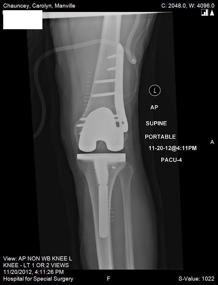
Carolyn Chauncey
Stamford, CT
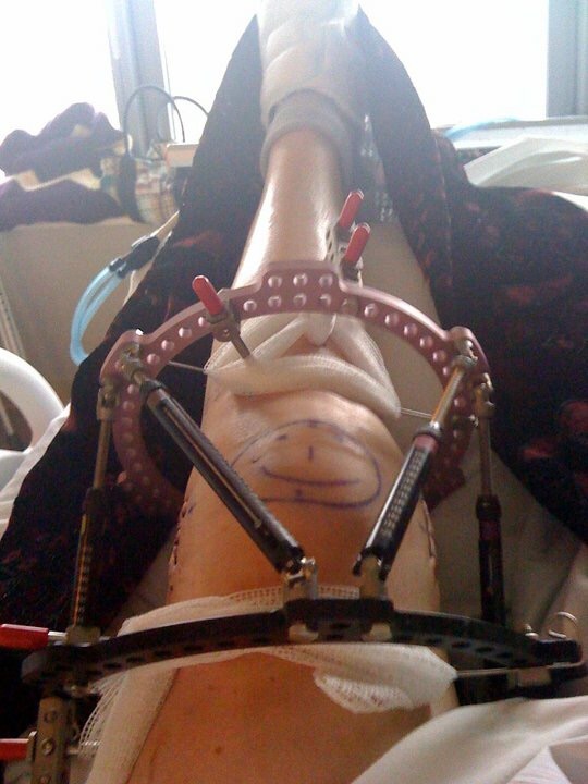
Broken Leg (Leg Fracture)
James Schwalbe
Englewood, NJ
ACL Tear (Torn ACL)
Joseph Franco
St. James, NY
Knee Pain Causes, Conditions and Treatments
Sherwin Slater
Monroe Township, NY
Kathryn Yurchuk
Plattekill, NY
Knee Replacement
Michael Bushell
Old Greenwich, CT
Shoulder Pain Causes, Conditions and Treatments
Joseph Murphy
Staten Island, NY
Knee Pain Causes, Conditions and Treatments
Eric Johnson
Norwalk, CT
ACL Tear (Torn ACL)
Santa J Johnston
Chesapeake, VA
Knee Replacement
Bruce Dixon
Greenwich, CT
Knee Replacement
Richard DeLuca
Stony Point, NY
Dupuytren's Contracture
Meghan Marchica
New York, NY
ACL Tear (Torn ACL)
Jane Jones
Suffern, NY
Knee Pain Causes, Conditions and Treatments
Tina Malizia
Seaside Park, NJ
Knee Arthritis
Richard del Bello
Westport, CT
Essence Carson
Paterson, NJ
ACL Tear (Torn ACL)